NIF in Focus: Inside the ARC Laser
September 19, 2019
"NIF in Focus" narrows in to explore important science and technology at the National Ignition Facility.
The Advanced Radiographic Capability (ARC) laser resides inside the giant laser system of the National Ignition Facility (NIF). An extremely large and powerful short-pulse laser, ARC operates in unison with NIF to give researchers both an unparalleled diagnostic tool and a very powerful energy source that can drive experiments investigating the frontiers of science.
The following figures taken from the animation above give a more in-depth description of how ARC works as a laser within a laser (Click each diagram to expand):
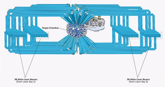
Figure 1. NIF's 192 high-energy, main laser beams each travel through their own dedicated beamline (shown in light blue). Two separate laser bays house these beamlines (96 beamlines per laser bay). However, all the main laser beams originate from a single low energy pulse of 1 billionth of a joule (1×10⁻⁹ J) that is eventually split and amplified to a total energy of about 4.8 megajoules (4.8×10⁶ J), an increase of nearly 5 quadrillion times. This figure picks up after the amplification with the main laser beams zooming down their beamlines at the speed of light to a target chamber that is 10 meters (33 feet) in diameter, the size of a small hot air balloon. When ARC is used during an experiment, it replaces the main laser beams in two of these beamlines.
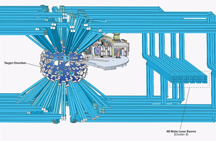
Figure 2. Zooming in reveals more details of the target chamber. Eventually, ARC and NIF’s main laser beams will arrive at the center of the target chamber. The circular ports around the chamber are typically where positioners and diagnostics are mounted. To simplify the diagram, they are not shown. All of the main laser beams, distributed evenly, enter the top and bottom of the target chamber through laser ports in groups of 4. The 96 main laser beams in each laser bay are arranged into two 48-beam clusters. These groupings and arrangements ensure every main laser beam has been properly amplified and timed to enter the target chamber at a precisely defined moment.
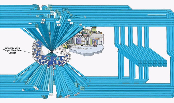
Figure 3. A section of the target chamber wall is cut away to show how all the main laser beams simultaneously come together at the target chamber center. The experimental target is positioned at the point where they all converge. As shown in this experiment, only the main laser beams are firing. ARC has not fired but soon will.
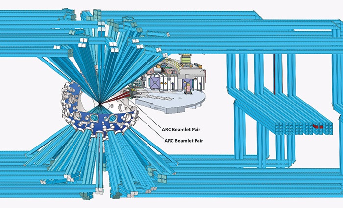
Figure 4. This diagram shows ARC firing into the target chamber during an experiment. ARC uses 2 beamlines (highlighted in red). The main laser beams are deactivated in these 2 beamlines when ARC is used. Only one laser fires through the shared beamlines for any given experiment.
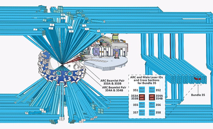
Figure 5. Another important distinction between ARC and the main laser beams is that each beamline used by ARC contains 2 separate, side-by-side ARC beamlets. The callout shows the laser identifications and configuration for Bundle 35, which is a group of 8 beamlines, including the 2 used by ARC. A total of 4 ARC beamlets come from these 2 beamlines.
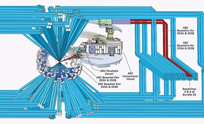
Figure 6. With some beamlines removed, the ARC beamlets' path toward the target chamber is more visible. Using a process called chirped pulse amplification (CPA), ARC produces extremely short duration, high-energy pulses without damaging or destroying NIF's optics. The pulse shape of each ARC beamlet is initially stretched, spreading out the laser energy over time. The beamlets are then amplified in energy to a maximum safe level. Essentially, the beamlets are broadened to provide more room to safely pack in more energy. After this amplification and before entering the target chamber, the beamlets enter the ARC compressor vessel and then the ARC parabola vessel.
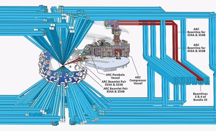
Figure 7. Inside the ARC compressor vessel each beamlet hits a series of four diffraction gratings, the final phase of the CPA process. These diffraction gratings progressively squeeze together the ARC beamlets, making them very short in pulse duration. During experiments, the pulse duration can range from about 1.3 to 38 picoseconds (a picosecond, or ps, is 1 trillionth of a second) and each beamlet can be fired at an energy of up to 1 kJ. More energy over a shorter time increases the power (power = energy/time) of the beamlets, which now have safely passed through most of NIF’s optics. The ARC parabola vessel contains parabolic mirrors that focus the beamlets to the size of a pinpoint at the center of the target chamber.
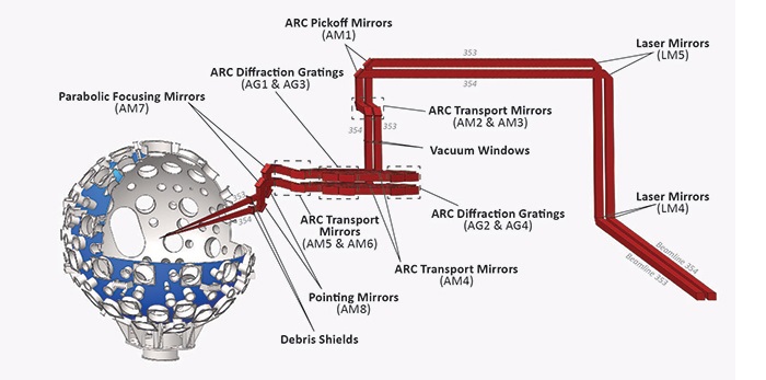
Figure 8. In this last figure, only the ARC beamlines and target chamber remain. Identified are all the mirrors used to direct the beamlets through the beamlines and the positions of the diffraction gratings and parabolic focusing mirrors. Also shown are the pointing mirrors, which, depending on the requirements of an experiment, can point all four beamlets at the same spot, each pair at different spots, or each beamlet at different spots.
Visit the links below to find out more.
To see an animation showing how ARC works, go to NIF | How the ARC Laser Works.
To learn about the chirped pulse amplification process that allows the ARC laser to operate, go to “Nobelist’s Invention Helped Spark LLNL’s Short-pulse Laser Breakthroughs.”
For more info about the ARC laser and its recent experiments, go to Advanced Radiographic Capability (ARC) Laser.
Diagrams by Paul Bloom and James Wickboldt.
—Dan Linehan
Follow us on Twitter: @lasers_llnl


